Difference between revisions of "File:BrainWindow020.png"
From MIPAV
(The region under the cursor (in the upper middle section of the image) displays the structural MRI that is otherwise hidden from view by the functional MRI. The structural MRI shows different data of the same brain than the functional MRI making the curso) |
|||
| Line 1: | Line 1: | ||
| − | The region under the cursor (in the upper middle section of the image) displays the | + | The region under the cursor (in the upper middle section of the image) displays the functional MRI that is otherwise hidden from view by the structural MRI. The structural MRI shows different data of the same subject compared to the functional MRI, making the cursor-window display a different view of the same brain. |
Latest revision as of 20:03, 23 March 2009
The region under the cursor (in the upper middle section of the image) displays the functional MRI that is otherwise hidden from view by the structural MRI. The structural MRI shows different data of the same subject compared to the functional MRI, making the cursor-window display a different view of the same brain.
File history
Click on a date/time to view the file as it appeared at that time.
| Date/Time | Thumbnail | Dimensions | User | Comment | |
|---|---|---|---|---|---|
| current | 19:46, 23 March 2009 | 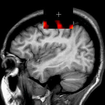 | <widthheight> (23 KB) | Mccreedy (Talk | contribs) | The region under the cursor (in the upper middle section of the image) displays the structural MRI that is otherwise hidden from view by the functional MRI. The structural MRI shows different data of the same brain than the functional MRI making the curso |
- You cannot overwrite this file.
File usage
The following page links to this file: