Image overlays and composites
MIPAV provides the ability to view two data sets at once. Simply stated, one image sits on top of the other. Using the MIPAV controls to favor one image over the other (image blending or fusion), the user can control what the user will see.
We have created a video using MIPAV to demonstrate the overlaying of image volumes and image blending.
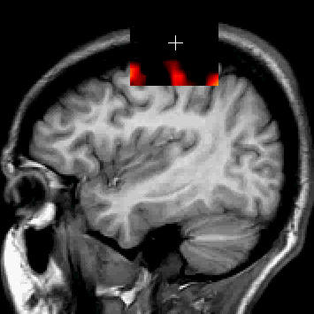
Image B Cursor Window
Two scans of the same patient can displayed as seperate images, but it may be more useful to the researcher to view the images together. This view -- where the underlying image is all or partly invisible except when viewed by the window, allows a researcher to focus on one image, while picking the second image data when need be. The image at right displays the sMRI data displayed with the window cursor, and is displayed over the functional MRI. Otherwise, the sMRI is invisible.
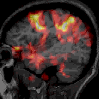
Image B Alpha-blending
The alpha-blending of two images loaded into the same MIPAV frame can be adjusted to that only one or the other of them is visible, or anywhere in between. Different lookup tables (LUTs) can be applied to each data set to make differences in the two images more apparent.

